Anatomy education continues to evolve as medical schools seek more flexible and sustainable teaching methods. Procuring and storing organic specimens requires significant budget allocation year after year. Exposure to harsh preservation chemicals like formaldehyde poses continuous health risks to both students and educators.
Institutions are increasingly seeking safer, scalable alternatives to meet growing enrollment numbers. Digital technologies offer a practical and highly effective solution. These systems replicate complex human structures using exact anatomical data. They provide risk-free, repeatable environments for intricate procedural practice.
Overcoming Traditional Specimen Shortages
Relying solely on physical cadavers creates significant bottlenecks in modern medical education. Organic tissues degrade rapidly after repeated classroom handling and dissection. This physical wear limits the frequency of hands-on practice available to each trainee. An accidental cut by a student can permanently ruin a delicate anatomical structure for the entire class.
Modern facilities mitigate these issues by implementing advanced digital alternatives. A high-resolution anatomy table provides infinite repeatability without physical degradation. Students can perform virtual incisions repeatedly until they achieve procedural mastery. This technological shift significantly reduces recurring disposal fees and eliminates the need for specialized ventilation systems.
Physical specimens also require dedicated storage facilities with rigorous temperature controls. Replacing these requirements with software-based solutions frees up valuable campus real estate. Universities can expand open lab hours because the digital environment requires no hazardous material supervision.
Core Applications in Medical Curricula
Advanced digital systems serve multiple departments within a comprehensive university. They are not limited strictly to standard systemic observation. The software platforms feature diverse learning modules to cover an entire medical curriculum. These modules include regional structures, embryology, and cross-sectional imaging.
Instructors utilize these comprehensive systems for diverse teaching scenarios. During a lecture, educators can strip away virtual tissue layers instantly with a simple touch. This reveals the complex spatial relationships among deep organ systems and skeletal structures. Trainees can use the platform to review anatomical structures before simulation-based clinical training. They can analyze specific pathological cases before entering a live clinical setting.
Data-Driven Precision and Structural Accuracy
The value of any educational technology relies heavily on its high structural integrity. Standard textbooks and static plastic models lack the depth needed for real-world surgical preparation. Modern digital platforms utilize authentic human tomographic imaging data as their foundation. This extensive data ensures highly accurate structural restoration and clear tissue layering.
Users observe intricate systems with a precision ranging from 0.1mm to 1.0mm. This precise level of detail exposes subtle nerves and micro-vessels clearly. The software reconstructs these fine details into a fully manipulative three-dimensional format. Learners can isolate, rotate, and magnify over 6,000 specific anatomical structures on the screen.
Enhancing Clinical Training and Diagnostic Skills
Effective medical training requires integration with real-world clinical diagnostic scenarios. Advanced digital platforms incorporate vast libraries of diagnostic imaging directly into the system. Students can access more than 1,700 CT and MRI scans seamlessly integrated with the 3D models. This allows trainees to compare radiological scans directly with corresponding tomographic specimen images.
The inclusion of a digital histological slice library adds another layer of instructional value. Users interact with the screen just as they would with a physical clinical microscope. They can adjust magnifications instantly across multiple objective lens settings. This meets multi-level teaching needs from macroscopic observation down to fine cellular structures.
Improving Doctor-Patient Communication
Clinical professionals often face challenges when explaining complex diagnoses to their patients. Traditional two-dimensional scans usually confuse individuals without a formal medical background. Three-dimensional visualizations bridge this critical communication gap effectively.
Practitioners use digital models to illustrate specific medical conditions clearly and visually. They can show patients exactly where a tumor is located or how a bone fracture impacts surrounding tissue. This visual clarity helps patients fully understand their proposed treatment plans. Informed patients often experience reduced anxiety regarding their upcoming procedures. Clear communication also streamlines the informed consent process in busy clinical environments.
Comprehensive Software and Hardware Integration
To support this level of detailed instruction, universities turn to specialized engineering manufacturers. Educational technology providers such as DIGIHUMAN offer integrated digital anatomy platforms for medical education. Their core platforms support multi-mode displays and semantic association for complex medical terminology.
Furthermore, these modern ecosystems often expand beyond standard flat screens. Institutions incorporate medical 3D models alongside medical teaching products based on VR and AR technologies. This multi-platform approach accommodates different learning styles and spatial comprehension needs.
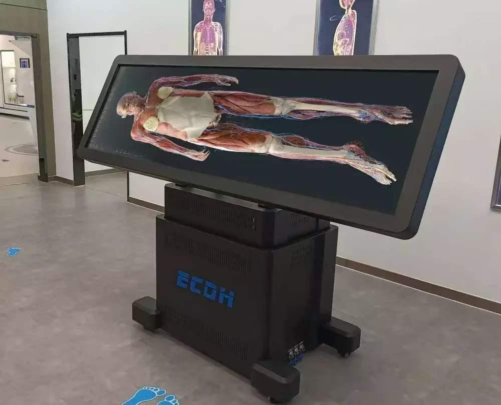
The core software integrates smoothly with standard teaching courseware and presentation files. Educators use intuitive multi-touch commands for quick navigation between different lecture topics. The hardware itself supports seamless large-group interaction. Standard professional setups feature large 88-inch high-definition displays that can tilt 90 degrees. This flexibility ensures optimal viewing angles for entire student cohorts during a live demonstration.
A Sustainable Upgrade for Educational Facilities
Transitioning to digital dissection curricula represents a strategic financial upgrade for medical institutions. The initial hardware investment provides years of utility without the recurring costs of organic tissue procurement. Software updates continually introduce new clinical cases and pathological data to the system. Trainees can add their own 3D annotations directly onto the digital models for future review.
Medical schools must prepare their graduates for a highly technology-driven healthcare landscape. Training them on interactive 3D interfaces mirrors the advanced diagnostic tools utilized in modern hospitals. A reliable anatomy table helps students develop a deep, spatial understanding of the human body. This strong foundational knowledge directly translates to safer clinical practices and improved patient outcomes.
